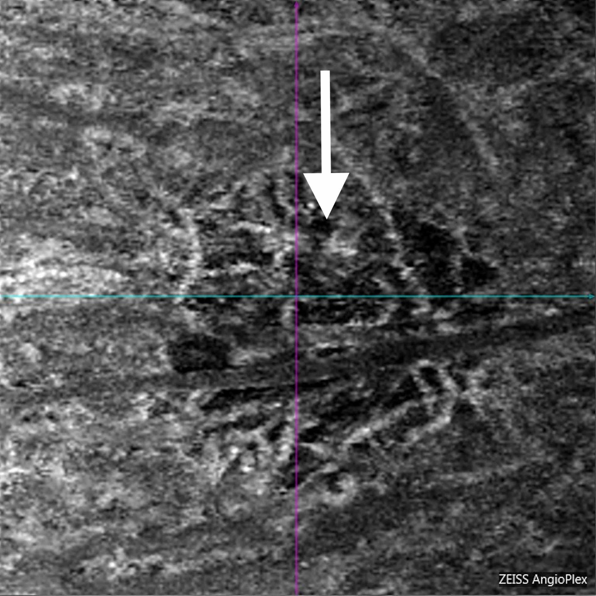

As the scanning beams enter the eye, areas with no movement will always reflect light the same way. OCTA works by scanning the same location on the retina multiple times consecutively. With the introduction of OCTA, clinicians now have a fast, noninvasive method to visualize retinal microvascular perfusion.

Additionally, indocyanine green dye is contraindicated in pregnant patients, or patients with kidney disease 2. The injected dye can sometimes cause adverse reactions including nausea, vomiting, and in rare occasions, anaphylaxis possibly leading to death 1. They are invasive, time consuming, and require a skilled photographer. Despite their great clinical utility, these procedures are not without drawbacks. Until now, clinical visualization of retinal vasculature has been limited to indocyanine green angiography (ICGA) and fluorescein angiography (FA), both of which require contrast dye injections. This allows for visualization of blood flow in the retina without the need for intravenous injection of a contrast dye.

OCTA isolates microvascular circulation from OCT image data by specialized acquisition and processing techniques. Recently, there has been great excitement in the development of a new OCT imaging technique called OCT angiography (OCTA). It has since become one of the most important ophthalmic diagnostic tools, with an expanding number of applications that aid eye care providers in diagnosing and monitoring eye disease. Optical coherence tomography (OCT) has revolutionized eye care by providing a fast, noninvasive method to visualize ocular structures in vivo since its development in 1991. Sha has a continued commitment to furthering the field of optometry in both her research and in private practice. Sha maintains an active role in the optometry community and is a part of the American Academy of Optometry, American Optometric Association, California Optometric Association, and the Santa Clara County Optometric Society, where she acts as the Legislative Events Liaison. Sha also practices private eye care in the Bay Area, CA. Sha works as a research coordinator on new medical devices relied upon by the optometric community. Dr. Sha became an Optometric Consultant at Carl Zeiss Meditec, Inc. As a consultant, Dr. Sha earned her Doctor of Optometry degree from the University of California, Berkeley, School of Optometry, where she was a part of the Beta Sigma Kappa Optometric Honor Society. Patricia Sha, OD earned her Bachelor of Science in Biochemistry and Cell Biology from the University of California, San Diego.
OPTICAL COHERENCE TOMOGRAPHY ANGIOGRAPHY FREE
In his free time, he enjoys spending time with friends and family, watching movies, and recording music. He is also an active member of his local optometric society. Chen is a member of the American Optometric Association, the California Optometric Association, and American Academy of Optometry. He also serves as a clinical research consultant to Carl Zeiss Meditec for advanced product and prototype development, and has worked extensively in the area of OCT angiography. He currently resides in the San Francisco Bay Area, and works in a private practice in Santa Clara, CA. Chen graduated from UC Berkeley School of Optometry.


 0 kommentar(er)
